
The human eye allows us to see the world around us. Its intricate anatomy processes light and images so our brain can interpret what we view. When part of this delicate system fails, vision loss and even blindness can occur.
The eye sits in a protective, cone-shaped cavity called the orbit or socket. It measures about an inch across. Layers of fat in the orbit cushion the eye and enable it to move easily. Six muscles control eye motion.
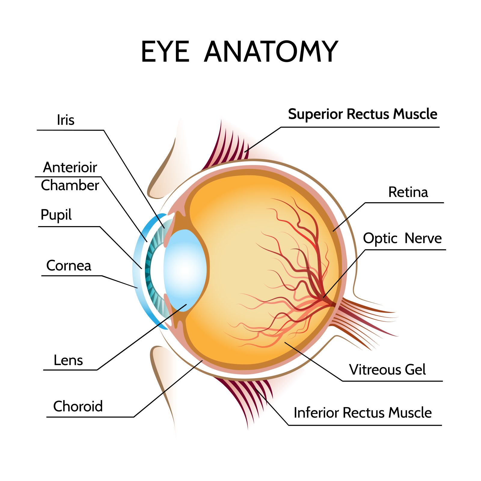
Key parts of the eye anatomy include:
Cornea – The cornea is the outermost, clear layer of the eye. The main function of the cornea is the refraction of light and focusing. Around 60 to 70% of the focusing ability of eyes is due to the cornea. The rest comes from the lens of the eye. Cornea quickly heals from minor abrasions. LASIK surgery reshapes this part of the eye to fix nearsightedness, farsightedness, astigmatism, and presbyopia.
Lens – Situated behind the iris, the lens is responsible for the other 30-40% of the eye’s focusing ability. With age, the lens hardens which decreases the accommodation ability of the eye causing one to need bifocals or reading glasses. It can also become opacified also known as a “cataract”.
Iris – The colored part of the eye. It controls light levels entering the eye by adjusting pupil size.
Pupil – The dark hole at the eye’s center that regulates light intake by dilating and constricting.
Retina – Light-sensitive nerve tissue lining the eye’s rear interior transmits images to the brain like a camera film. Focused light rays must hit the center of the eye “the macula” for perfect vision.
Optic Nerve – Carries electric signals encoding images from retina to brain.
Sclera – The eye’s white outer layer giving shape and protection.
Multiple parts work together for sight. When any part malfunctions, vision loss or even legal blindness can occur. Understanding eye anatomy reveals how delicate but vital human vision is.
Categories